Techniques
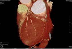
Cardiovascular MRI and CT
We offer a toolbox of novel quantitative cardiac and vascular MRI and CT techniques measuring global and regional cardiac function, microvascular perfusion, and structure as well as angiography and 2D and 4D flow measurements.
Publications
https://www.ncbi.nlm.nih.gov/pubmed/?term=vogel-claussen+cardiac
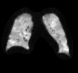
19F MRI
MRI of fluorinated tracers is an emerging approach for monitoring of chronic obstructive lung diseases. The negligible endogenous appearance of 19F in the human body and its application even at thermal polarization due to its gyromagnetic ratio close to hydrogen facilitate 19F MRI as a sensitive tool with potential clinical translation
References
Gutberlet M; Kaireit TF; Voskrebenzev A; Kern AL; Obert A; Wacker F; Hohlfeld J; Vogel-Claussen J, Repeatability of regional lung ventilation quantification using fluorinated (19F) gas magnetic resonance imaging. Acad Radiol. 2018 Nov 22. pii: S1076-6332(18)30485-9. doi: 10.1016/j.acra.2018.10.021
Kaireit TF, Gutberlet M, Voskrebenzev A, Freise J, Welte T, Hohlfeld JM, Wacker F, Vogel-Claussen J. Comparison of quantitative regional ventilation-weighted fourier decomposition MRI with dynamic fluorinated gas washout MRI and lung function testing in COPD patients. J Magn Reson Imaging. 2018 Jun;47(6):1534-1541.
Gutberlet M, Kaireit T, Voskrebenzev A, Lasch F, Freise J, Welte T, Wacker F, Hohlfeld JM, Vogel-Claussen J, Free-breathing dynamic fluorinated gas MRI for mapping of regional lung ventilation in COPD patients. Radiology. 2018 Mar;286(3):1040-1051
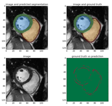
Machine Learning
Machine learning made significant progress during the last years and shows continuous exponential growth in many fields of research. Provided with sufficient training data, new deep learning algorithms are able to outperform humans at repetitive tasks. The access to high quality data in combination with exceptional computational power and expertise allows us to create highly automated image-analysis pipelines for effective and reproducible workflow.
References
Winther HB, Hundt C, Schmidt B, Czerner C, Bauersachs J, Wacker F, Vogel-Claussen J. ν-net: Deep Learning for Generalized Biventricular Mass and Function Parameters Using Multicenter Cardiac MRI Data. JACC Cardiovasc Imaging. 2018 Jul;11(7):1036-1038.
Winther HB, Hundt C, Schmidt B, Czerner C, Bauersachs J, Wacker F, Vogel-Claussen J. ν-net: Deep Learning for Generalized Biventricular Cardiac Mass and Function Parameters, 2017, arXiv:1706.04397 [cs.CV]
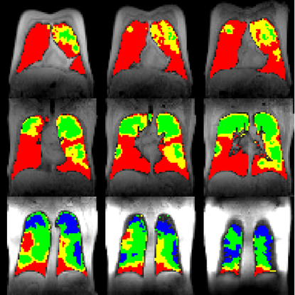
PREFUL
Phase-Resolved Functional Lung Imaging (PREFUL) is a non-invasive, patient friendly MRI technique to assess pulmonary ventilation (V) and perfusion (Q). The images are acquired during free breathing without any contrast agent using widely available sequence and hardware technology. Refined post-processing allows to extract a wide range of sensitive parameters, including dynamic information. In summary, PREFUL is a powerful ventilation-perfusion scan without ionizing-radiation.
References
Voskrebenzev A, Gutberlet M, Klimeš F, Kaireit TF, Schönfeld C, Rotärmel A, Wacker F, et al. Feasibility of quantitative regional ventilation and perfusion mapping with phase-resolved functional lung (PREFUL) MRI in healthy volunteers and COPD, CTEPH, and CF patients. Magn Reson Med. 2018;79(4):2306-2314. doi: 10.1002/mrm.26893


Quantitative Lung CT
We offer a toolbox of quantitative lung CT measurements including emphysema quantification and parametric response mapping of the lung. In close collaboration with Prof. Dr. med. Hoen-oh Shin, we develop novel biomarkers for regional lung function.
References
https://www.ncbi.nlm.nih.gov/pubmed/?term=vogel-claussen+lung+computed+tomography

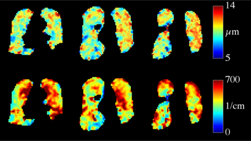
129Xe MRI
MRI of hyperpolarized 129Xe has proven to be a highly versatile tool for the quantitative assessment of lung function and microstructure. By the unique property of xenon among known MRI tracer gases of being soluble in tissues and blood, 129Xe MRI renders entirely new applications in research as well as the clinic possible.
References
Kern AL, Gutberlet M, Voskrebenzev A, Klimes F, Rotärmel A, Wacker F, Hohlfeld JM, Vogel-Claussen J. Mapping of regional lung microstructural parameters using hyperpolarized 129Xe dissolved-phase MRI in healthy volunteers and patients with chronic obstructive pulmonary disease. Magn Reson Med. 2019;81(4):2360–2373 doi: 10.1002/mrm.27559
Kern AL, Gutberlet M, Qing K, Voskrebenzev A, Klimes F, Kaireit TF, Czerner C, Biller H, Wacker F, Ruppert K, Hohlfeld JM, Vogel-Claussen J. Regional investigation of lung function and microstructure parameters by localized 129Xe chemical shift saturation recovery and dissolved-phase imaging: A reproducibility study. Magn Reson Med. 2019;81(1):13–24 doi: 10.1002/mrm.27407
Kern AL, Vogel-Claussen J: Hyperpolarized gas MRI in pulmonology. Br J Radiol. 2018;91:20170647 doi: 10.1259/bjr.20170647